Want to learn more about surgical guide planning?
Discover how digital workflows can improve your implant success rates.
Table of Contents
- The Impression Is the Foundation — And It's Where Most Cases Fail
- What We Actually Receive: The Data Problem
- Real Numbers from Our Lab
- Why Alginate Fails for Surgical Guide Applications
- 1. Dimensional Instability (Syneresis and Imbibition)
- 2. Tear Strength and Detail Reproduction
- 3. The Pour-and-Scan Pipeline
- Why Digital Impressions Win: It's Not Just About Accuracy
- Direct Surface Capture
- Scan Bodies and Implant-Level Registration
- Immediate Quality Verification
- The Critical Alignment Step: CBCT-to-STL Registration
- How Registration Works
- What Happens with Poor STL Data
- What About PVS? The Middle Ground
- The Economics of Going Digital
- Our Recommendations: What to Send Us
- Ideal Submission (Fastest, Most Accurate)
- Acceptable (With Caveats)
- Not Recommended
- How to Check Your STL Quality Before Submitting
- The Bottom Line
- Accuracy Comparison: Intraoral Scanner vs Alginate
- When Analog Impressions Still Make Sense
- Hybrid Workflow: Combining Both
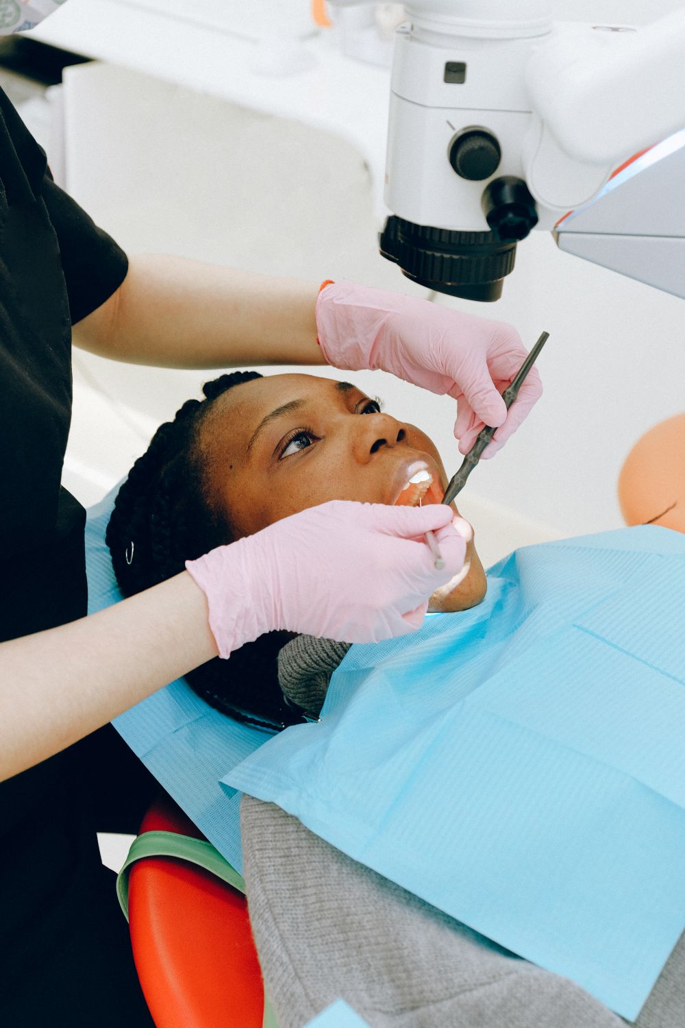
The Impression Is the Foundation — And It's Where Most Cases Fail
Every surgical guide is only as good as the surface data it's built on. You can have a perfect CBCT scan, flawless implant planning in coDiagnostiX or Blue Sky Plan, and a precisely calibrated 3D printer — but if your impression data is inaccurate, the guide won't seat, the occlusion won't match, and the implant position will deviate from plan.
A 3D-printed template that fits over the patient's teeth or tissue and directs drill placement during implant surgery. It transfers the digital treatment plan into precise physical drill positions.
A 3D imaging technique that captures the jaw, teeth, and bone structure in a single rotational scan. It produces DICOM files used for implant planning, nerve mapping, and surgical guide design.
After designing surgical guides from over 4,000 submitted impression files, we can say this with certainty: the single most common cause of guide inaccuracy is poor impression quality. And in 2026, that almost always means an analog impression.
---
What We Actually Receive: The Data Problem
When clinicians submit cases for surgical guide design, we receive one of three types of surface data:
- STL files from intraoral scanners (TRIOS 3/4/5, Primescan, Medit i700, iTero) — ~72% of submissions
- PVS (polyvinyl siloxane) impressions scanned by the lab — ~18% of submissions
- Alginate impressions scanned or poured into stone models — ~10% of submissions
The quality difference is not subtle. It's measurable.
Real Numbers from Our Lab
| Parameter | Intraoral STL | Lab-Scanned PVS | Scanned Alginate Model |
|---|---|---|---|
| Mean surface deviation | 32 µm (±12) | 67 µm (±28) | 142 µm (±55) |
| Rejection rate (unusable data) | 3.1% | 8.7% | 23.4% |
| Requires rescan/reimpression | 2.2% | 6.3% | 19.8% |
| Average turnaround delay | 0 days | +0.5 days | +1.8 days |
Translation: alginate-based workflows are 7x more likely to require a redo than digital impressions, and they add nearly 2 days to your case turnaround on average.
---
Why Alginate Fails for Surgical Guide Applications
Alginate (irreversible hydrocolloid) was designed for study models and provisional restorations — not for micron-level precision work. Here's why it's fundamentally incompatible with guided implant surgery:
An implant placement technique that uses a physical surgical guide to direct drills and implants to positions planned in 3D software. It improves accuracy and reduces surgical risks compared to freehand placement.
1. Dimensional Instability (Syneresis and Imbibition)
Alginate begins losing water immediately upon removal from the mouth. Within 15 minutes, linear shrinkage reaches 0.3–0.5%. On a full-arch impression, that translates to 0.15–0.25mm of cumulative distortion across the arch.
For a single-tooth restoration, that's acceptable. For a surgical guide that needs <100µm of seating accuracy? It's a guaranteed misfit.
2. Tear Strength and Detail Reproduction
Alginate has a tear strength of approximately 350–700 g/cm compared to PVS at 2,000–5,000 g/cm. In interproximal areas and around healing abutments, alginate tears during removal — creating voids in exactly the areas where guide seating depends on accurate surface reproduction.
3. The Pour-and-Scan Pipeline
Even if the alginate impression is perfect at chairside, it then goes through:
- Stone pouring (introduces 0.05–0.1% expansion)
- Model trimming (introduces operator variability)
- Lab scanning (introduces scanner error: ~15–25 µm)
Each step compounds error. By the time we receive the digital model, we're working with data that has been degraded through 4 separate error sources.
---
Why Digital Impressions Win: It's Not Just About Accuracy
Direct Surface Capture
An intraoral scanner captures the tooth surface directly — no impression material, no stone model, no lab scanner intermediate. The STL file we receive is one step removed from the patient's anatomy.
A 3D surface mesh file format used in dental CAD/CAM. Intraoral scanners produce STL files that capture tooth and gingival surfaces for surgical guide fitting.
Modern scanners achieve trueness of 20–40 µm for full-arch scans (validated by The Journal of Prosthetic Dentistry, 2024). For partial arch scans (implant sites with 2–4 adjacent teeth), trueness drops to 12–20 µm — well within the tolerance required for guided surgery accuracy.
Scan Bodies and Implant-Level Registration
For cases involving existing implants or healing abutments, intraoral scanners with scan bodies provide direct implant-position data. This is impossible with alginate — the scan body geometry is too complex for hydrocolloid reproduction.
We strongly recommend:
- TRIOS 3/4/5 — excellent scan body recognition, native AI-assisted stitching
- Primescan — highest single-capture depth (20mm), very fast
- Medit i700/i900 — best price-to-accuracy ratio for practices scaling into digital workflows
Immediate Quality Verification
With a digital scan, the clinician can verify completeness chairside — checking for holes, artifacts, and marginal capture before the patient leaves. With alginate, you discover problems hours or days later, requiring a recall.
---
The Critical Alignment Step: CBCT-to-STL Registration
This is where digital impression accuracy has its biggest impact on guide quality and where most clinicians underestimate the consequences.
How Registration Works
Surgical guide planning requires superimposition of two datasets:
- CBCT data — bone, roots, nerve canals, sinuses
- Surface scan (STL) — teeth, soft tissue, prosthetic surfaces
The software matches the STL surface to the visible tooth crowns in the CBCT using a best-fit algorithm (typically ICP — Iterative Closest Point). The quality of this registration determines everything downstream.
What Happens with Poor STL Data
When the STL surface is distorted (from alginate shrinkage, pour expansion, or scan artifacts), the ICP algorithm produces a suboptimal fit. The registration may look acceptable on screen, but the RMS error (root mean square of point-to-point distances) is elevated.
Our quality threshold:
- <80 µm RMS — excellent registration, proceed with planning
- 80–150 µm RMS — marginal, may need manual adjustment
- >150 µm RMS — reject, request new scan
From our data:
- STL from intraoral scanner: 94% achieve <80 µm on first attempt
- STL from PVS model scan: 78% achieve <80 µm on first attempt
- STL from alginate model: 51% achieve <80 µm on first attempt
This means nearly half of alginate-derived cases require either manual registration correction or a completely new impression. Each correction introduces operator-dependent error.
---
What About PVS? The Middle Ground
Polyvinyl siloxane (addition silicone) impressions are significantly better than alginate:
- Dimensional stability: stable for weeks (vs. minutes for alginate)
- Tear strength: 4–7x higher than alginate
- Detail reproduction: 20 µm (vs. 50+ µm for alginate)
Our recommendation: if you don't have an intraoral scanner yet, PVS is acceptable for surgical guide work — but it still introduces the pour-scan pipeline error. Consider it a transitional solution, not a long-term workflow.
The Economics of Going Digital
For practices placing 5+ implants per month, the ROI on an intraoral scanner is typically 8–14 months:
- Material savings: ~€8–12 per impression (alginate + tray vs. disposable sleeve)
- Lab scanning fees eliminated: €15–25 per model
- Rescan/recall rate reduction: from ~20% to ~3%
- Turnaround time: 1–2 days faster per case
- Patient experience: no gagging, no taste, instant verification
---
Our Recommendations: What to Send Us
Based on our experience designing guides from every type of impression data:
Ideal Submission (Fastest, Most Accurate)
- Intraoral STL scan in .STL or .PLY format
- Scan body scan if implant-level registration is needed
- Opposing arch scan + bite registration scan
- Export at maximum resolution (do not reduce mesh)
Acceptable (With Caveats)
- PVS impression → lab desktop scan → STL file
- Ensure the lab uses a calibrated scanner (3Shape D2000, Medit T710, or equivalent)
- Trim model minimally — we need the full arch for registration
Not Recommended
- Alginate impressions — too many variables, too high rejection rate
- Photographs or 2D radiographs as sole diagnostic data
- Low-resolution STL exports (some scanners default to "light" export mode)
---
How to Check Your STL Quality Before Submitting
A quick quality check before sending files to your surgical guide design partner:
- Open the STL in MeshLab or 3D viewer — look for holes, spikes, or floating fragments
- Check file size — a full-arch STL should be 15–40 MB. Under 5 MB suggests the mesh is decimated
- Inspect the scan margin — ensure at least 2–3 teeth adjacent to the implant site are fully captured
- Verify bite registration — intercuspation should be stable, no "floating" contact points
- Confirm orientation — most software expects upper jaw facing up (this saves us processing time)
---
The Bottom Line
The shift from analog to digital impressions isn't a luxury — it's a clinical necessity for any practice offering guided implant surgery. Alginate impressions introduce cumulative errors that directly compromise surgical guide fit, CBCT-STL registration accuracy, and ultimately implant placement precision.
If you're still sending alginate impressions for surgical guide cases, you're paying more (in remakes and delays) for worse outcomes. The data is clear.
> Ready to go digital?
> Send us your intraoral STL scan and CBCT — we'll deliver a precision surgical guide designed in 24–48 hours. First case review is free.
Accuracy Comparison: Intraoral Scanner vs Alginate
Multiple clinical studies have demonstrated that intraoral scanners achieve trueness within 20-50 microns for single arch scans, compared to 50-100+ microns for alginate impressions. This difference becomes critical in surgical guide design where every micron affects implant placement accuracy.
For full-arch cases, however, the accuracy gap narrows. Intraoral scanners can accumulate errors across the arch, resulting in deviations of 100-200 microns at the distal ends. In these cases, a combination of segmented digital scans stitched with reference markers can improve accuracy.
The key advantage of digital impressions for surgical guides is the seamless integration with CAD software. STL files from intraoral scanners can be directly imported into guide design software, eliminating the need for model scanning and its associated errors.
When Analog Impressions Still Make Sense
Despite the clear advantages of digital workflows, analog impressions remain relevant in specific clinical scenarios:
Limited mouth opening: Patients with restricted opening may not accommodate the scanner tip, making traditional impression trays more practical.
Subgingival margin capture: Deep subgingival preparations can be challenging for optical scanners. Polyvinyl siloxane (PVS) materials can capture details below the gingival margin more reliably.
Cost considerations: For clinics without intraoral scanners, investing in a scanner solely for surgical guide cases may not be economically justified when outsourced scanning services are available.
Hybrid Workflow: Combining Both
Many advanced clinics adopt a hybrid approach: digital impressions for the working arch and CT-based virtual models for the opposing arch and bone anatomy. This combination leverages the surface accuracy of optical scanning with the volumetric data of CBCT scans.
The surgical guide design process benefits most when both datasets are accurately aligned using fiducial markers or anatomical landmarks. Modern CAD platforms support automatic registration of STL and DICOM data, creating a unified virtual patient model.
Digital Imaging and Communications in Medicine — the universal file format for medical imaging. CBCT scanners produce DICOM files that are imported into planning software for 3D reconstruction.
Ready to streamline your surgical guide workflow?
Join 200+ dental professionals who trust SurgicalGuide.Pro for precision planning.


