Want to learn more about surgical guide planning?
Discover how digital workflows can improve your implant success rates.
Table of Contents
Endodontic microsurgery — commonly known as apicoectomy — demands pinpoint accuracy. The surgeon must locate the root apex, remove precisely 3mm of infected tissue, and seal the canal retrograde, all while preserving adjacent anatomical structures.
Traditional approaches rely on periapical radiographs and clinical experience. But when the root is curved, the apex is close to the mental nerve, or the tooth has multiple canals, a 2D image simply is not enough.
A 3D-planned apical resection guide solves this problem by translating CBCT data into a physical template that shows the surgeon exactly where to cut.
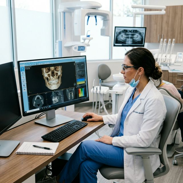
A 3D imaging technique that captures the jaw, teeth, and bone structure in a single rotational scan. It produces DICOM files used for implant planning, nerve mapping, and surgical guide design.
How an Apical Resection Guide Works
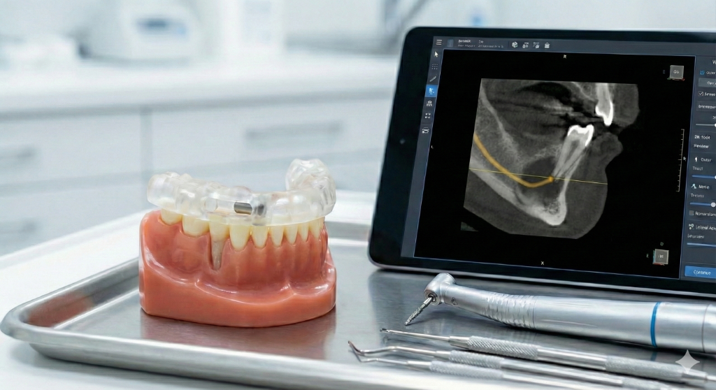
The guide is a 3D-printed template, similar in concept to an implant surgical guide. It fits over the patient's teeth and contains a precisely positioned channel or window that directs the surgical bur to the exact location of the root apex.
A 3D-printed template that fits over the patient's teeth or tissue and directs drill placement during implant surgery. It transfers the digital treatment plan into precise physical drill positions.
The design process follows a familiar digital workflow:
- CBCT scan reveals the 3D root anatomy, including curves, accessory canals, and proximity to vital structures
- Digital planning identifies the target osteotomy site and optimal approach angle
- Guide design positions a drill channel or access window aligned to the planned resection
- 3D printing produces the guide in biocompatible resin
- Surgery proceeds through the guide with controlled, predictable access
Clinical Advantages
- Reduced osteotomy size — the surgeon accesses only the necessary area, preserving cortical bone
- Faster apex location — no exploratory drilling or repeated verification with radiographs
- Nerve protection — critical when operating near the inferior alveolar nerve or mental foramen
- Consistent resection depth — the guide controls not just position, but also the angle of approach
- Shorter procedures — typical surgical time is reduced by 20-40%
When to Use a Guided Approach
An apical resection guide is most valuable when:
- The root apex is not palpable through bone
- Multiple roots require resection in a single session
- The tooth is adjacent to the mental foramen or inferior alveolar canal
- Previous apicoectomy has failed and re-intervention is planned
- The referring dentist or patient demands minimally invasive surgery
Our Workflow at SurgicalGuide.Pro
You upload the CBCT data, specify which tooth requires resection, and our team designs a precision guide. We deliver the print-ready STL file within 48 hours, ready for your in-office printer or local lab.
A 3D surface mesh file format used in dental CAD/CAM. Intraoral scanners produce STL files that capture tooth and gingival surfaces for surgical guide fitting.
FAQ
Can you design guides for multi-rooted teeth?
Yes. We routinely design apical resection guides for molars with two or three separate root apices, each with its own guided access channel.
What is the minimum CBCT resolution needed?
We recommend a voxel size of 0.2mm or smaller for optimal root visualization. Most modern CBCT units meet this requirement.
Is the guide reusable?
No. Each guide is patient-specific and designed for single use, following biocompatibility and sterilization standards.
---
Planning an apicoectomy?
Upload your CBCT and receive a precision resection guide.
Create an Order on SurgicalGuide.pro
Ordering an Apical Resection Guide Design Online
The entire apical resection guide workflow can be completed remotely. You do not need to visit a lab or mail physical models.
The online process:
- Upload your CBCT (DICOM) and intraoral scan (STL) through our secure platform
- Specify the target tooth and resection depth
- Receive a 3D treatment plan with the guide design within 2-3 business days
- Review the plan in your browser — no software installation required
- Download the print-ready STL file and print locally or through your lab
Online guide design eliminates geographical limitations. Whether your clinic is in Berlin, Dubai, or São Paulo, you receive the same professional design quality.
Ready to streamline your surgical guide workflow?
Join 200+ dental professionals who trust SurgicalGuide.Pro for precision planning.